
IHuman Case Study Case Study 60year Old Female Shortness Of Breath Evaluation Of Dyspnea. Class 6512 Week #7
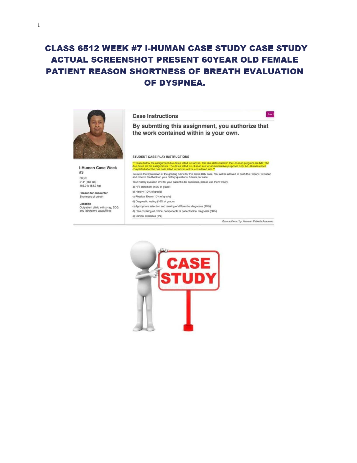
IHuman Case Study Case Study 60year Old Female Shortness Of Breath Evaluation Of Dyspnea. Class 6512 Week #7
Shortness of Breath: Comprehensive iHuman Case Study
Here's a complete human case study based on your request:
Case Study: Shortness of Breath
Patient Information
• Age: 60 years old
• Sex: Female
• Height: 5'4" (163 cm)
• Weight: 183.0 lbs (83.3 kg)
• BMI: 32.4 (Obese Class I)
Chief Complaint
"I've been feeling more short of breath over the last few weeks."
5
History of Present Illness (HPI)
The patient is a 60-year-old woman who presents with progressive shortness of breath over the past 3 weeks. She notes that the dyspnea is worse with exertion, such as climbing stairs or walking for more than a block, and has gradually become noticeable even at rest. She denies chest pain but reports mild fatigue and occasional wheezing. She has not had fever, chills, night sweats, or recent infections. She reports some orthopnea and mild ankle swelling in the evenings. No history of trauma or recent travel.
She has no known history of asthma or COPD but mentions she smoked for about 15 years in her 20s and 30s (approximately 15 pack-years). She quit over 20 years ago.
Past Medical History
• Hypertension (diagnosed 10 years ago)
• Hyperlipidemia
• Type 2 Diabetes Mellitus (controlled with metformin)
• Obesity
• Former smoker (quit 20+ years ago)
Medications
• Metformin 1000 mg BID
• Lisinopril 20 mg daily
• Atorvastatin 40 mg nightly
• Aspirin 81 mg daily
Allergies
• No known drug allergies (NKDA)
6
Family History
• Father died of myocardial infarction at age 65
• Mother with hypertension and type 2 diabetes
Social History
• Lives with husband
• Retired schoolteacher
• No alcohol or drug use
• Ex-smoker (15 pack-years, quit >20 years ago)
• Sedentary lifestyle
Review of Systems
• General: Fatigue, mild unintentional weight gain (5 lbs in 3 months)
• Cardiac: No chest pain, palpitations, or syncope
• Respiratory: Dyspnea, orthopnea, occasional wheezing, no cough
• GI: No nausea, vomiting, or abdominal pain
• MSK: No muscle weakness or joint pain
• Neuro: No headaches or dizziness
7
Physical Examination (PE)
Vital Signs:
• BP: 148/86 mmHg
• HR: 92 bpm
• RR: 20/min
• Temp: 98.6°F (37°C)
• SpO₂: 92% on room air
General: Alert, oriented, mild respiratory distress at rest
Neck: No JVD
Lungs: Bibasilar crackles, mild expiratory wheeze Heart: Regular rhythm, no murmurs, rubs, or gallops Abdomen: Soft, non-tender, no hepatosplenomegaly Extremities: 1+ bilateral ankle edema
Skin: No cyanosis or rash
Initial Workup / Tests
• CBC: WNL
• BMP: Mildly elevated glucose (145 mg/dL), otherwise WNL
• BNP: 380 pg/mL (elevated)
• CXR: Cardiomegaly, mild pulmonary vascular congestion
• EKG: Normal sinus rhythm, no ST changes
• Echocardiogram: EF 45%, mild LV hypertrophy, diastolic dysfunction
• Spirometry: Mild obstruction, FEV1/FVC slightly decreased
8
Differential Diagnosis
1. Heart Failure with Preserved Ejection Fraction (HFpEF)
2. Chronic Obstructive Pulmonary Disease (COPD) – mild/reactive component
3. Obesity Hypoventilation Syndrome
4. Pulmonary Hypertension
5. Anemia
6. Deconditioning
Diagnosis
• Primary: Heart Failure with Preserved Ejection Fraction (HFpEF)
• Contributing: Mild obstructive lung disease (former smoker), obesity-related
hypoventilation
Management Plan
Lifestyle and Risk Factor Modification
• Sodium restriction, daily weights, fluid restriction
• Encourage gradual physical activity (e.g., cardiac rehab)
• Weight loss counseling: Refer to nutritionist
• Smoking cessation reinforcement (though quit >20 years ago)
Pharmacologic
• Initiate diuretic (e.g., furosemide 20 mg daily) for volume overload
• Continue ACE inhibitor (lisinopril) for blood pressure and cardiac remodeling
• Consider beta-blocker (e.g., carvedilol) if HR remains elevated
• Continue statin and metformin
• Consider low-dose spironolactone if persistent symptoms
Monitoring
• Follow-up in 1–2 weeks to reassess weight, BP, symptoms, and labs
• Monitor renal function and electrolytes with diuretic therapy
9
• Annual echocardiogram for EF monitoring
• Pulmonary function tests annually
Education
• Educate on HF warning signs: increased SOB, weight gain, swelling
• Importance of daily weights, medication adherence
• Discuss advanced care planning if disease progresses
Would you like this case formatted for use in an EMR template or teaching slide deck?
Here is a case study of a 60-year-old female presenting with shortness of breath, structured for educational or clinical purposes:
Human Case Study: Shortness of Breath
Patient Profile:
• Name: Mrs. Linda Garcia
• Age: 60
• Sex: Female
• Occupation: Office administrator
• Medical History:
o Hypertension (10 years)
o Hyperlipidemia
o Obesity (BMI 33)
o No smoking history
o No known drug allergies
Reason for Encounter:
"I’ve been short of breath for the past few days, and it’s getting worse."
10
History of Present Illness:
Mrs. Garcia reports the onset of shortness of breath 4 days ago, which began during mild exertion and has progressively worsened. Today, she felt breathless even while walking to the bathroom. She also mentions a dry cough and mild chest tightness but denies chest pain, fever, leg swelling, or recent travel. She feels fatigued and has been sleeping propped up on two pillows for the past two nights.
Review of Systems:
• Respiratory: Dyspnea on exertion, orthopnea, dry cough
• Cardiovascular: Chest tightness, no palpitations or syncope
• General: Fatigue, no fever or chills
• Other systems: Unremarkable
Vital Signs:
• BP: 148/88 mmHg
• HR: 104 bpm
• RR: 24 breaths/min
• Temp: 37.0°C
• SpO₂: 91% on room air
• BMI: 33 kg/m²
Physical Examination:
• General: Mild respiratory distress, alert and oriented
• Chest: Bibasilar crackles, no wheezing
• Heart: Regular rhythm, no murmurs
• Neck: Jugular venous distention (JVD) present
• Extremities: Mild bilateral ankle edema, no cyanosis
• Abdomen & Neuro: Normal
Initial Investigations:
• ECG: Sinus tachycardia, no ischemic changes
• CXR: Cardiomegaly with mild pulmonary congestion
• BNP: Elevated (820 pg/mL)
• CBC & CMP: Within normal limits
• Echocardiogram: Reduced ejection fraction (~40%), left ventricular hypertrophy
• D-dimer: Negative
• COVID-19 test: Negative